The temporomandibular joint (TMJ) is the hinge and sliding mechanism that links your lower jaw (mandible) to the skull. Unlike a simple hinge, each TMJ can move up and down, forward and backward, and side to side — a combination that allows chewing, speaking and wide opening motions like yawning. A thin, shock-absorbing disc sits between the jaw’s rounded condyle and the temporal bone, helping the joint move smoothly under load.
Because the TMJ combines rotational and translational movement in a small, highly coordinated space, it is more complex than many other joints in the body. Muscles, ligaments and dental contacts all influence how the joint functions, so a problem in one element can affect the whole system. That interconnectedness is one reason TMJ conditions often present with varied and overlapping symptoms.
Understanding this anatomy helps patients and clinicians recognize why treatment needs to be targeted and individualized. Rather than assuming a single cause, clinicians consider the joint’s structural components, muscle behavior and how the bite (occlusion) interacts with everyday habits like chewing and clenching.
TMJ disorders can show up in many ways, and symptoms often extend beyond the jaw itself. Common complaints include jaw pain or stiffness, limited opening or closing, and noises such as popping, clicking or grinding when the mouth moves. These sensory signs often accompany difficulty chewing and occasional episodes of the jaw feeling “stuck” or locking in an open or closed position.
Because the TMJ sits close to the ear, patients frequently report ear-related symptoms like aching, fullness or even tinnitus (ringing). Headaches, particularly around the temples, and tension or pain in the neck and upper shoulders are also frequent companions of TMJ dysfunction, reflecting how muscular tension in one area can radiate to nearby structures.
Symptoms can be intermittent or persistent, mild or disabling. Some patients notice flare-ups tied to stress, poor sleep habits, or episodes of jaw overuse such as excessive gum chewing or teeth grinding. Tracking patterns — what makes symptoms better or worse — provides valuable clues for diagnosis and management.
Clinicians generally group TMJ disorders into three broad categories to guide evaluation and treatment. Myofascial pain involves the muscles that control the jaw: chronic tension, spasms or fatigue in these muscles can generate significant discomfort. Internal derangement refers to mechanical problems within the joint, such as disc displacement, dislocation, or trauma to the condyle. Degenerative conditions include arthritic changes that erode joint surfaces over time.
Risk factors that raise the likelihood of developing TMJ issues include long-term teeth grinding (bruxism), previous jaw injury, certain inflammatory or degenerative arthritides, and repetitive strain from occupational or recreational activities. Psychological factors such as prolonged stress can also play a role by increasing muscle tension and clenching behavior.
Often, more than one contributing factor is present. For example, muscle dysfunction may coexist with a displaced disc or early arthritic change, which is why a thorough, multi-factorial approach to assessment produces the most reliable results and more predictable treatment responses.
Identifying the primary drivers of a patient’s symptoms — muscle versus joint structural change versus degenerative process — helps clinicians select the least invasive and most effective interventions first, reserving more complex procedures for persistent or severe cases.
Evaluation begins with a detailed clinical history and careful physical examination. A clinician will assess jaw range of motion, listen for joint sounds, palpate the muscles and joint for tenderness, and observe how the teeth meet. Understanding everyday activities and habits, including sleep behavior and stressors, is an important part of the diagnostic picture.
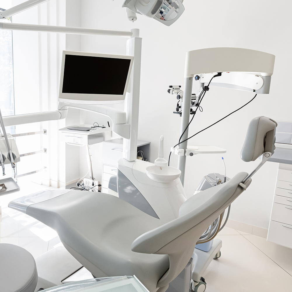
When needed, imaging supports the clinical exam. Panoramic or cone-beam radiography can show bony changes and condylar position, while MRI is the preferred tool for visualizing soft tissues such as the articular disc. These tests are ordered selectively based on clinical findings and the suspected underlying issue rather than used routinely for every patient.
Additional assessments may include bite analysis and evaluation of occlusion to identify contacts that place abnormal strain on the joint. In some cases, referral to or collaboration with orofacial pain specialists, physical therapists, or ENT providers helps clarify complex presentations and coordinate care across disciplines.
Treatment typically follows a conservative, staged approach. For many patients, initial management focuses on reversible, low-risk measures: behavioral changes (avoiding hard or chewy foods, limiting wide yawning), sleep hygiene and stress-reduction techniques, and temporary use of a removable oral appliance to reduce clenching forces. These interventions aim to reduce inflammation, relax muscles and improve joint comfort while diagnostic evaluation continues.
Physical therapy and guided home exercises can reestablish normal range of motion and address muscle imbalances through stretching, posture correction and manual techniques. When inflammation or severe muscle spasm is present, short-term pharmacologic support under a clinician’s direction can provide relief and allow participation in rehabilitative exercises.
For cases that don’t respond to conservative care, targeted procedures may be considered. These can include intra-articular therapies to address inflammation, occlusal adjustments when dental contacts significantly contribute to symptoms, or referral for orthodontic or prosthodontic solutions when bite relationships require correction. Surgery is rarely the first choice and is reserved for specific structural problems unresponsive to other treatments.
Throughout care, successful outcomes depend on clear communication between patient and clinician, careful monitoring of symptoms, and flexible treatment planning that prioritizes function, comfort and long-term joint health.
Summary — If you suspect a temporomandibular disorder, early assessment improves the chance of resolving symptoms with conservative measures. The temporomandibular joint’s complexity means evaluation should consider muscles, joint structures and dental contacts together. Our team at Suss Dental Group has experience evaluating TMJ conditions and coordinating appropriate care. Contact us to learn more about diagnosis and treatment options and to discuss what approach may be best for your situation.


A temporomandibular joint (TMJ) disorder refers to a group of conditions that affect the jaw joint and the muscles that control jaw movement. These disorders can involve the joint’s soft tissues, the articular disc, the bony condyle or the surrounding muscles and ligaments, and they often produce pain, altered motion or mechanical noises.
Because the TMJ permits both rotational and sliding movement, dysfunction can disrupt chewing, speaking and wide opening motions. Clinicians approach TMJ disorders as multifactorial problems that require assessment of muscle behavior, joint structure and how dental contacts influence joint loading.
Common symptoms include jaw pain or stiffness, limited mouth opening, and audible sounds such as clicking, popping or grinding when the jaw moves. Many patients also report difficulty chewing, episodes of the jaw feeling stuck or locked, and intermittent flare-ups that vary with activity or stress.
Because the TMJ is adjacent to the ear and cranial muscles, symptoms frequently extend beyond the joint and include ear fullness, aching, tinnitus, temple headaches and tension in the neck and shoulders. Tracking when symptoms worsen or improve helps clinicians identify contributing behaviors and tailor treatment.
TMJ disorders arise from a combination of factors rather than a single cause; common contributors include long-term teeth grinding or clenching (bruxism), jaw trauma, degenerative joint disease and internal derangement such as disc displacement. Repetitive strain from occupation or hobbies, abnormal bite relationships and inflammatory conditions can also increase risk.
Psychological stress and poor sleep habits may exacerbate muscle tension and clenching behavior, making symptoms more likely or more severe. Often patients present with overlapping issues, for example muscle dysfunction alongside early joint degeneration, which is why a comprehensive assessment is important.
Evaluation begins with a thorough medical and dental history followed by a focused physical exam that assesses jaw range of motion, joint sounds, muscle tenderness and how the teeth meet. Clinicians also observe functional movements, palpate masticatory muscles and check for signs of occlusal interference that may place abnormal strain on the joint.
When indicated, imaging is used selectively: panoramic or cone-beam radiographs for bony changes and MRI to visualize the articular disc and soft tissues. At Suss Dental Group our clinicians coordinate findings with potential referrals to orofacial pain specialists, physical therapists or ENT providers when the presentation is complex or multidisciplinary care is needed.
Initial management emphasizes reversible, low-risk interventions such as behavioral modifications, a soft or modified diet, and strategies to reduce clenching and parafunctional habits. Short-term use of a removable oral appliance can reduce joint loading while home care, sleep hygiene and stress-reduction techniques address contributing behaviors.
Physical therapy and structured home exercises help restore range of motion and relieve muscle imbalances through stretching, posture correction and manual techniques. Clinicians may also recommend short-term pharmacologic support or intra-articular therapy in selected cases to control inflammation and enable active rehabilitation.
Occlusal splints or night guards are typically recommended when clenching or bruxism is a significant factor in a patient’s symptoms and when reducing tooth-to-tooth contact will likely lower joint strain. These removable appliances are designed to redistribute occlusal forces, protect dental surfaces and permit muscle relaxation during sleep or high-stress periods.
Splints are a conservative, reversible option and are often used as part of a broader management plan that includes habit modification and physical therapy. Their design and wear schedule are individualized based on examination findings and the patient’s functional needs.
Yes. Physical therapy is an effective component of TMJ care for many patients because it targets muscle tightness, postural imbalances and restricted joint mobility that contribute to symptoms. Interventions may include manual therapy, therapeutic exercises, education on posture and ergonomics, and modalities to reduce pain and inflammation.
Guided home exercise programs teach patients how to maintain improvements and reduce recurrences, while coordinated care between the dental clinician and a skilled physical therapist improves treatment consistency. Progress is monitored and exercise programs are adapted as range of motion and muscle control improve.
Imaging or referral is considered when the clinical exam suggests structural joint problems, when symptoms are severe or persistent despite conservative care, or when there are red flags such as progressive joint degeneration or a history of trauma. MRI is the best modality for visualizing disc position and soft tissues, while cone-beam or panoramic radiographs evaluate bony anatomy.
Referral to orofacial pain specialists, oral and maxillofacial surgeons, physical therapists or ENT and rheumatology colleagues is appropriate when multidisciplinary input is needed. These collaborations help clarify complex cases and guide targeted therapies beyond basic conservative measures.
Surgical interventions are reserved for specific structural problems that do not respond to conservative care, such as certain forms of internal derangement or advanced degenerative change. Options range from minimally invasive procedures like arthrocentesis and arthroscopy to open joint surgery for cases where direct structural repair or reconstruction is necessary.
Because surgery carries risks and recovery time, clinicians exhaust nonsurgical treatments and obtain appropriate imaging and specialist opinions before recommending operative approaches. When surgery is indicated, careful planning and clear communication about goals and realistic outcomes are essential.
Preventive measures focus on minimizing joint stress and addressing modifiable contributors: avoid hard or chewy foods, limit wide yawning and nail or pen biting, and adopt stress-management techniques to reduce clenching. Maintaining good posture, especially during prolonged computer or phone use, helps reduce strain on neck and jaw muscles that can precipitate symptoms.
Regular dental checkups let clinicians monitor occlusion and dental wear patterns that may affect the joint, and early evaluation of new symptoms improves the likelihood that conservative care will be effective. If symptoms persist or worsen, seek a professional assessment so a tailored plan can protect joint function and comfort over the long term.
Ready to schedule your next dental appointment or have questions about our services?
Contacting Suss Dental Group is easy! Our friendly staff is available to assist you with scheduling appointments, answering inquiries about treatment options, and addressing any concerns you may have. Whether you prefer to give us a call, send us an email, or fill out our convenient online contact form, we're here to help. Don't wait to take the first step towards achieving the smile of your dreams – reach out to us today and discover the difference personalized dental care can make.