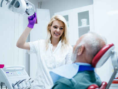
Oral cancer screening is a straightforward step that can be built into a routine dental exam, and it plays a quiet but vital role in preserving long-term health. Many early signs of oral cancer are subtle and painless, which means they can be missed outside of a clinical setting. When clinicians include a focused screening in regular checkups, they improve the chances of catching abnormalities at an early, more treatable stage.
Screening is not a one-size-fits-all procedure — it’s a preventive habit. For people with known risk factors or who notice persistent changes in their mouth, more frequent checks are often advised. Even for those without obvious risks, a professional visual and tactile exam can identify things the average person might not detect or might underestimate.
Making screening routine helps normalize the process and reduces anxiety for patients. Framing it as a standard part of oral health care emphasizes prevention and early detection rather than waiting for symptoms to worsen. That proactive mindset is central to how our practice approaches comprehensive dental wellness.
During a screening, clinicians systematically inspect the entire oral cavity and related structures. This includes the lips, tongue (both top and underside), gums, inner cheeks, floor of the mouth, hard palate, and the oropharynx as far as can be viewed. The neck and surrounding lymph nodes are also palpated to detect swelling or firmness that could indicate an underlying problem.
Practitioners look for a range of visual and physical clues: persistent ulcers, white or red patches, lumps, thickened areas of tissue, and any surface changes that do not resolve within a couple of weeks. The texture and mobility of tissue are just as important as color or appearance; a lesion that doesn’t move freely under the finger is worth closer attention.
Because oral tissue varies greatly between individuals, clinicians compare suspicious findings against the patient’s baseline. What may be normal for one person could be unusual for another, so screening is both observational and contextual — informed by medical history, lifestyle factors, and any recent changes reported by the patient.
Certain behaviors and exposures raise the probability of developing cancers in the mouth and throat. These include tobacco use, heavy alcohol consumption, prolonged ultraviolet exposure to the lips, and infection with high‑risk strains of human papillomavirus (HPV). Age and prior radiation treatment to the head and neck can also play a role. Knowing which factors apply to an individual helps tailor screening frequency and follow-up recommendations.
Early warning signs are often easy to miss because they can feel harmless at first: small sores that don’t heal, areas of persistent irritation, new lumps or thickening, or unexplained numbness. Changes in speech, swallowing, or a persistent sore throat can also be meaningful. Reporting these symptoms sooner rather than later gives clinicians a better chance of intervening early.
Open communication between patient and provider about lifestyle, sexual health, and prior treatments contributes to a more effective screening strategy. Patients who understand their personal risk profile are better positioned to recognize concerning changes and to collaborate on an appropriate monitoring schedule.
The central element of screening remains a careful visual and tactile exam performed by a trained clinician. In many practices, that foundational assessment is supplemented by technology designed to enhance visibility or highlight abnormal tissue. Examples include adjunctive light-based aids and intraoral cameras that magnify and document suspicious areas for closer study over time.
These tools are intended to assist the clinician, not replace clinical judgment. Imaging and magnification can make subtle differences more apparent and create a record to compare at future visits. When findings are unclear or persistent, clinicians may recommend further diagnostic steps such as biopsy or referral to a specialist for definitive evaluation.
Clinicians also consider the whole-patient context when choosing tools and next steps — balancing thoroughness with the patient’s comfort and medical history. The goal is to use available resources efficiently to clarify whether an abnormality is benign, warrants monitoring, or needs immediate intervention.
If a screening identifies an area of concern, the next step is a focused plan for diagnostic clarification. In some cases, the clinician will monitor the spot for a short interval to see if it resolves on its own. If tissue changes persist, a biopsy — the removal of a small sample for laboratory analysis — may be recommended to determine whether cancerous or precancerous cells are present.
Referral pathways are an important part of responsible screening. When specialized evaluation is needed, the clinician will coordinate with oral surgeons, otolaryngologists, or oncologists to ensure timely assessment. These coordinated steps help reduce delays between detection and diagnosis, which is important for achieving the best possible outcomes.
Supportive care and clear communication are essential during this process. Clinicians will explain the findings, describe possible next steps, and answer questions about what to expect. Keeping patients informed and involved in decisions helps reduce uncertainty and keeps the focus on effective, compassionate care.
Summary and next steps
Oral cancer screening is a practical, evidence‑based element of comprehensive dental care that emphasizes early detection and patient education. By combining careful clinical exams with appropriate use of modern tools and timely follow-up, clinicians can significantly improve the likelihood of identifying problems at an early stage. Suss Dental Group integrates screening into routine visits so patients benefit from steady, preventive oversight.
If you have questions about oral cancer screening or would like to learn more about how it fits into your routine dental care, please contact us for more information.


An oral cancer screening is a focused clinical exam designed to identify early signs of cancer or precancerous changes in the mouth and throat. It combines a systematic visual inspection with gentle palpation of tissues to detect abnormalities that might not be noticeable to a patient. The goal is early detection so that any necessary diagnostic follow-up can occur promptly and with better potential outcomes.
Screening is a preventive service that can be completed during a routine dental visit and typically takes only a few minutes. It looks beyond obvious cavities and gum disease to evaluate the lips, tongue, floor of the mouth, cheeks, palate and neck. When findings are uncertain, clinicians document them and recommend monitoring or further diagnostic steps rather than immediate alarm.
Many early signs of oral cancer are painless and easy to miss without a professional exam, so including screening in routine dental care increases the chance of catching problems early. Dentists and hygienists are trained to recognize subtle changes in color, texture, or mobility of oral tissues that merit attention. Early detection improves treatment options and the likelihood of successful outcomes.
Making screening a regular part of dental checkups also normalizes the process and reduces patient anxiety about unknown findings. When clinicians discuss risk factors and symptoms during the visit, patients are better informed about what to watch for between appointments. This collaborative approach helps patients and providers create an appropriate monitoring plan based on individual needs.
A comprehensive screening inspects the entire oral cavity and related structures, including the lips, top and underside of the tongue, inner cheeks, gums, hard palate and floor of the mouth. Clinicians also view as much of the oropharynx as possible and palpate the neck and lymph nodes to detect unusual firmness or swelling. Both visual clues, such as persistent red or white patches, and physical signs, such as lumps or immobile tissue, are important.
The examiner compares any suspicious findings with the patient’s normal baseline and medical history to determine their significance. Areas that do not resolve within two weeks or that change over time typically receive closer evaluation. Documentation and intraoral photography are often used to track changes between visits.
Several factors increase the likelihood of developing oral and oropharyngeal cancers, including tobacco use, heavy alcohol consumption and infection with high‑risk strains of human papillomavirus (HPV). Age and prior radiation therapy to the head and neck also raise risk, and prolonged sun exposure to the lips contributes in certain cases. These elements are considered when clinicians recommend screening frequency and follow-up.
People with a history of significant tobacco or alcohol use, a weakened immune system, or a family history of cancer should be particularly vigilant about regular screenings. However, oral cancer can occur in individuals without obvious risk factors, so routine screening remains valuable for most adult patients. Open discussion of lifestyle and medical history allows clinicians to tailor monitoring appropriately.
For most adults, an oral cancer screening is performed at every routine dental exam, which commonly occurs every six months, but the recommended interval may vary based on individual risk. Patients with known risk factors, persistent oral sores, or a history of oral lesions may need more frequent checks. Your clinician will use your medical history and current findings to suggest an appropriate schedule.
If a clinician documents an area of concern that is being monitored, they will set a specific timeline for re‑evaluation and clearly communicate what signs should prompt an earlier appointment. Consistent follow-up reduces the risk of delayed diagnosis and ensures that changes are assessed in context. Staying engaged with regular visits gives clinicians the best chance to spot meaningful trends early.
The core of screening remains a careful visual and tactile exam, but adjunctive aids such as intraoral cameras and light‑based devices can improve visibility and documentation. Intraoral cameras magnify and photograph suspicious areas for more detailed assessment and comparison over time, while specialized light tools may help highlight abnormal tissue patterns. These technologies are meant to assist clinical judgment rather than replace it.
When a finding is unclear, clinicians may recommend imaging, a biopsy, or referral to a specialist for definitive evaluation. Using technology appropriately helps clarify whether an area should be observed, sampled, or treated. Decisions about which tools to use are made with consideration for the patient’s comfort, history and the clinical picture.
If an abnormality is detected, clinicians first determine whether short‑term observation is appropriate or whether immediate diagnostic steps are warranted. For lesions that do not resolve or that appear suspicious, a biopsy may be recommended to provide a tissue diagnosis. In other situations, timely referral to an oral surgeon, otolaryngologist or oncologist ensures specialized evaluation and coordination of care.
Throughout this process, clear communication and supportive information are key to reducing uncertainty for the patient. Clinicians explain findings, outline likely next steps and answer questions about what to expect during diagnostic procedures. Prompt, coordinated follow‑up helps minimize delays between detection and any necessary treatment.
Yes — oral cancer can often be detected at an earlier, more treatable stage when regular screening is performed and suspicious changes are investigated promptly. Early detection typically allows for less extensive treatment, better functional outcomes and a higher probability of long‑term survival. Detecting precancerous conditions also provides an opportunity for interventions that reduce progression to invasive disease.
Patients benefit from increased treatment options and a lower likelihood of complex reconstructive procedures when cancers are found early. In addition, monitoring and preventive counseling can address modifiable risk factors to reduce the chance of recurrence or new lesions. An emphasis on early identification is a cornerstone of effective oral health care.
Reducing risk involves addressing known contributors such as tobacco and heavy alcohol use, protecting the lips from excessive sun exposure and getting vaccinated against HPV when appropriate. Maintaining good oral hygiene, attending regular dental visits and reporting persistent mouth changes promptly also strengthen prevention efforts. Your clinician can offer personalized guidance based on your medical history and lifestyle.
Behavioral changes, including quitting smoking and limiting alcohol, have the strongest evidence for lowering oral cancer risk over time. Regular dental screenings and timely treatment of suspicious lesions add another layer of protection by enabling early intervention. Combining risk reduction with routine care provides the best overall strategy for protection.
Suss Dental Group incorporates a focused oral cancer screening into standard dental exams so patients receive consistent preventive oversight at their visits. The screening includes a systematic visual and tactile exam of all oral structures and palpation of the neck, and clinicians document findings to track any changes over time. When technology such as an intraoral camera is useful, it is used to magnify and record areas of concern for clearer follow‑up.
If an abnormality requires further evaluation, the practice coordinates timely next steps, which may include continued observation, biopsy or referral to a specialist for definitive diagnosis. Clinicians at the Bergenfield, New Jersey office prioritize clear communication so patients understand findings and recommended actions. This integrated approach emphasizes prevention, early detection and patient involvement in care decisions.
Ready to schedule your next dental appointment or have questions about our services?
Contacting Suss Dental Group is easy! Our friendly staff is available to assist you with scheduling appointments, answering inquiries about treatment options, and addressing any concerns you may have. Whether you prefer to give us a call, send us an email, or fill out our convenient online contact form, we're here to help. Don't wait to take the first step towards achieving the smile of your dreams – reach out to us today and discover the difference personalized dental care can make.