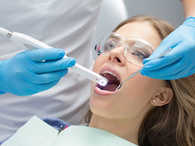
An intraoral camera is a compact, pen‑sized imaging device designed to capture detailed, full‑color views from inside the mouth. Using a tiny lens and integrated LED lighting, the camera takes high‑resolution stills and video that are displayed instantly on a nearby monitor. This real‑time visual feedback lets clinicians and patients see dental surfaces and soft tissues at magnifications that would be impossible with the naked eye.
Although small in size, intraoral cameras are engineered for clinical accuracy. The optics and illumination are optimized to reduce glare and highlight surface texture, enabling clear visualization of enamel defects, cracked teeth, margins of restorations, and the condition of the gums. Images can be adjusted for contrast and zoomed in without losing important detail, which helps clinicians evaluate subtle problems quickly and confidently.
Because the device captures both still photographs and short video sequences, it supports a range of clinical workflows—from quick documentation during a routine exam to more detailed imaging for treatment planning. The result is a precise, repeatable record of a patient’s oral condition that can be referenced over time or shared securely with other members of the dental care team.
Seeing is often the most persuasive form of explanation. When patients are shown clear images of their mouth during an exam, complex findings become easier to understand. Intraoral camera images help clinicians point out the exact location and nature of an issue—such as a hairline crack, recurrent decay around a filling, or inflamed tissue—so patients can make informed decisions about next steps.
These images also support patient education. Rather than relying on verbal descriptions alone, clinicians can walk patients through what they see on screen, explain the significance of different findings, and illustrate how proposed treatments will address the problem. Visual evidence encourages questions and ensures patients leave the appointment with a clearer grasp of their oral health.
At Suss Dental Group we incorporate intraoral imaging into routine visits so that records reflect both clinical observations and visual proof. Saved photographs become part of the patient chart, creating a transparent history that patients can review as their care advances.
Intraoral cameras are versatile diagnostic tools. They are excellent for spotting early enamel breakdown, marginal gaps around restorations, and surface stains that may indicate deeper structural problems. Because the camera provides magnified views, it can reveal fractures, craze lines, and other defects that might be missed during a cursory visual exam.
Beyond detection, intraoral images support treatment planning. Clear photographs help clinicians assess the extent of tissue involvement, determine whether conservative repair or a more extensive restoration is required, and document pre‑treatment conditions before procedures such as crowns, bridges, or implant restorations. In cases where multiple treatment options exist, images make it easier to explain tradeoffs and expected outcomes.
These images also play a role in monitoring clinical change over time. Comparative photographs taken across multiple visits allow clinicians to track progression or healing—useful for periodontal management, monitoring suspicious lesions, or evaluating the effectiveness of a given therapy.
While intraoral cameras complement other diagnostic tools—such as digital radiography and clinical probing—they often reveal surface and color details that X‑rays cannot. Using both types of information together produces a more complete diagnostic picture and improves treatment accuracy.
One of the strengths of intraoral imaging is how easily it fits into a digital practice. Images can be saved to the electronic health record, organized by date and tooth number, and referenced alongside radiographs and digital impressions. This centralized documentation simplifies case management and supports efficient collaboration when a case requires input from a specialist or a dental laboratory.
Modern intraoral cameras export common file formats that are compatible with practice management systems and lab communication platforms. Whether a clinician needs to send a photographic record to a laboratory technician for shade matching or attach images to a referral, the digital workflow reduces wait time and helps ensure accurate, consistent results.
Security and privacy are fundamental in handling digital records. Image files are stored and transferred under secure protocols consistent with standard healthcare data protections, and best practices in file naming and charting help maintain a clear, organized record for each patient.
As part of an integrated digital strategy, intraoral photography also supports patient engagement tools such as visual treatment summaries or secure patient portals where individuals can review their own images. This level of transparency fosters a collaborative relationship between the patient and the care team.
For patients, intraoral imaging is quick, comfortable, and noninvasive. During a standard exam the clinician will gently position the camera inside the mouth and move it to capture the areas of interest. Because the device is designed for oral use, it’s easy to maneuver into the positions needed to reveal posterior teeth, gum margins, and other hard‑to‑see surfaces.
Clinicians typically display images on a monitor so patients can watch in real time. This allows immediate discussion about findings and next steps. If needed, individual images are saved in the patient’s chart for future reference, progress tracking, or consultation with other providers. The process adds only a few minutes to a routine exam but delivers lasting benefit in the form of clearer diagnosis and documentation.
Strict infection control protocols are followed for every intraoral camera. Disposable sleeves or other protective barriers are used on the device, and all handling follows standard sterilization procedures to ensure patient safety. While intraoral cameras enhance visual assessment, they do not replace the need for complementary tests such as X‑rays when those are clinically indicated.
In summary, intraoral cameras are a practical, patient‑friendly technology that enhances clinical accuracy, improves communication, and strengthens the dental record. If you’d like to learn more about how intraoral imaging is used in our practice or what it might reveal during your next visit, please contact Suss Dental Group for more information.


An intraoral camera is a compact, pen-sized imaging device that captures high-resolution, full-color views of the inside of the mouth. It uses a small lens and integrated LED lighting to produce still photographs and short video that display in real time on a nearby monitor. The magnified images reveal surface detail and color that are difficult to see with the naked eye.
Although small, these cameras are engineered for clinical accuracy, with optics and illumination designed to reduce glare and highlight texture. Images can be adjusted for contrast and zoomed without losing key detail, which supports precise clinical evaluation. Captured photos become part of the patient record for documentation and longitudinal comparison.
An intraoral camera improves diagnosis by revealing subtle surface findings such as hairline cracks, marginal gaps around restorations, early enamel breakdown, and localized tissue changes. Magnified, high-resolution images allow clinicians to identify issues that might be missed during a cursory visual exam and to document findings accurately. The ability to capture both stills and video supports a range of clinical workflows from routine screening to more detailed assessments.
These images also enable comparison over time, which helps track progression or healing and informs decisions about intervention. Because intraoral imaging complements other diagnostic tools like digital radiography and probing, it contributes surface and color detail that X-rays cannot provide alone. Combining multiple data sources produces a more complete diagnostic picture and supports more confident treatment planning.
Yes. Intraoral imaging is noninvasive and generally quick and comfortable for most patients, adding only a few minutes to a standard exam. The device is designed for oral use and can be maneuvered easily to capture posterior teeth, gum margins, and other hard-to-see areas without causing discomfort. Disposable sleeves or protective barriers are used and handling follows strict infection control protocols.
Because the procedure is visual and does not involve radiation or invasive tools, it is appropriate for virtually all patients, including those who require gentle care. Clinicians will use the camera selectively when images will provide useful diagnostic or educational value. If complementary tests such as X-rays are needed, the clinician will explain why they are indicated.
Intraoral images play a key role in treatment planning by documenting pre-treatment conditions and clarifying the extent of tissue or structural involvement. Clear photographs help clinicians determine whether conservative repair or a more extensive restoration is appropriate and support decisions for crowns, bridges, veneers, or implant restorations. When multiple treatment options exist, images make it easier to explain tradeoffs and expected outcomes.
Photographs can also be used to communicate with specialists and dental laboratories, improving shade matching and ensuring consistent results. Organized, dated images tied to a patient chart create a reliable record that supports case coordination and follow-up. This documentation contributes to predictable care and helps manage complex restorative or cosmetic cases.
Intraoral cameras and digital radiographs serve different but complementary purposes in diagnosis. Cameras capture surface detail, color, and texture that X-rays cannot, while radiographs reveal internal structures such as roots and bone levels. Using both modalities together provides a more complete view of oral health and reduces the risk of missing findings that span surface and subsurface anatomy.
Clinical probing, visual inspection, radiographs, and intraoral photography each contribute unique information, and clinicians integrate these sources to form an accurate diagnosis. When an image suggests deeper involvement, the clinician can correlate photographic findings with radiographic evidence and clinical testing to determine the appropriate next steps. This multimodal approach improves diagnostic confidence and treatment outcomes.
During an exam a clinician will gently position the camera inside the mouth and move it to capture the areas of interest while images appear on a monitor. Patients can often view the images in real time, which allows immediate discussion about findings and next steps. The process is quick, typically adding only a few minutes to the appointment, and is performed with attention to comfort and safety.
Individual images are saved in the patient chart when helpful for documentation, progress tracking, or referral purposes. Clinicians explain what is being shown and how it relates to oral health so patients leave with a clearer understanding of their condition. If additional diagnostic tests are needed, the clinician will describe the rationale and how those tests complement the photographic record.
Intraoral images are integrated into digital practice workflows and saved to the electronic health record alongside radiographs and clinical notes. Modern systems organize images by date and tooth number, which simplifies case management and supports efficient collaboration when referrals or laboratory communication are required. File formats are chosen for compatibility with practice management systems and lab platforms.
Security and privacy are treated as fundamental priorities, with images stored and transferred under secure protocols consistent with standard healthcare data protections. Best practices in file naming, charting, and access controls help maintain patient confidentiality and an organized record for each patient. Patients may also have access to their images through secure patient portals when available.
Intraoral cameras are excellent for detecting surface signs that suggest cavities, cracks, or marginal failure around restorations, such as localized discoloration, loss of translucency, or visible fracture lines. These surface findings can indicate the need for further evaluation and guide targeted radiographs or clinical tests. Because cameras provide magnified views, they often reveal early or subtle changes that warrant closer inspection.
However, some forms of decay and internal cracks may not be visible on the surface and require radiographs or other diagnostic tools for confirmation. Clinicians use intraoral imaging as part of a comprehensive assessment and will combine photographic evidence with X-rays, probing, and clinical judgement to arrive at an accurate diagnosis. Images therefore guide, but do not replace, the full diagnostic process.
Visual evidence is often the most persuasive form of explanation, and intraoral images help patients understand the exact location and nature of a problem. Clinicians can walk patients through what is visible on screen, explain the significance of different findings, and illustrate how proposed treatments will address the issue. This transparency encourages questions and ensures patients leave with a clearer grasp of their oral health.
Images also make it easier to compare options and discuss expected outcomes, which supports shared decision making. When patients can see their own condition and review documented images over time, they are better equipped to participate in care choices and to follow recommended preventive or therapeutic steps. This collaborative approach strengthens trust and improves adherence.
Suss Dental Group integrates intraoral imaging into routine visits to support thorough exams, clear patient communication, and accurate documentation of oral health. Clinicians capture targeted photographs during exams and save images to the patient record so findings can be tracked across appointments or shared with specialists and laboratories when needed. This practice-level approach enhances case planning and continuity of care.
Within a modern digital workflow, intraoral images at the Bergenfield, New Jersey office are used alongside radiographs and digital impressions to produce coordinated treatment plans. Patients benefit from a transparent visual record that makes diagnoses easier to understand and follow up on. If you have questions about how intraoral imaging will be used during your visit, the team will explain the process and answer your concerns.
Ready to schedule your next dental appointment or have questions about our services?
Contacting Suss Dental Group is easy! Our friendly staff is available to assist you with scheduling appointments, answering inquiries about treatment options, and addressing any concerns you may have. Whether you prefer to give us a call, send us an email, or fill out our convenient online contact form, we're here to help. Don't wait to take the first step towards achieving the smile of your dreams – reach out to us today and discover the difference personalized dental care can make.