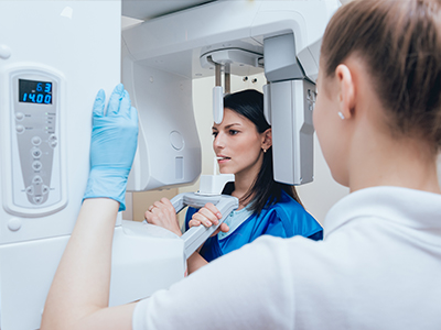
Digital radiography replaces traditional film with electronic sensors and computer processing to capture dental images. Instead of exposing film and developing it with chemicals, clinicians use compact sensors or phosphor plates that convert x‑ray information into digital files. Those files appear on a monitor within seconds, allowing immediate review and discussion during the same appointment. The change is more than convenience; it transforms how clinicians evaluate, document, and communicate about oral health.
At its core, the system consists of a detector, a workstation, and imaging software. When an x‑ray is taken, the sensor records data that the software translates into an image. Clinicians can then adjust brightness and contrast, zoom in on areas of interest, and apply measurement tools without re‑exposing the patient. This flexibility helps clinicians spot issues that might be harder to see on conventional film and streamlines clinical decision‑making.
For patients, the visible result is faster answers and a clearer explanation of findings. For the practice, digital radiography supports a higher standard of care by integrating imaging directly into the patient record and enabling more efficient coordination among specialists when collaborative treatment is needed.
Digital radiography is used across a wide range of dental services, from routine bitewings that detect early decay to periapical and panoramic images used for more complex assessments. Because images are available immediately, clinicians can confirm image quality at the chairside and retake a shot if needed — reducing the risk of missed diagnostic information. This immediacy improves workflow and keeps appointments on schedule without compromising diagnostic accuracy.
Integration with other digital tools makes radiography even more powerful. Images can be aligned with intraoral scans, digital impressions, and 3D planning software to support restorative work, implant placement, and orthodontic planning. The ability to overlay and compare different image sets helps clinicians monitor changes over time and craft treatment plans that are tailored to each patient’s anatomy and goals.
From a patient‑communication standpoint, digital images are an excellent teaching tool. Clinicians can enlarge an area, annotate concerns, and demonstrate how a proposed treatment will address a specific problem. This visual approach helps patients understand conditions and participate in informed decision‑making about their care.
One of the key advantages of digital radiography is reduced radiation exposure. Digital sensors are generally more sensitive to x‑rays than film, so they require a lower dose to produce a diagnostically useful image. While any medical imaging is performed only when clinically indicated, the lower exposure profile of digital systems contributes to safer routine care when radiographs are necessary.
Beyond dose reduction, the smaller size and design of modern sensors can improve comfort, particularly for patients with strong gag reflexes or limited mouth opening. Faster capture times also mean the patient spends less time holding a sensor in place. Clinicians still follow established safety protocols — including shielding and careful selection of imaging angles — to minimize exposure while maximizing diagnostic value.
Practices that adopt digital radiography typically combine these technical safety benefits with clear patient communication. By explaining why an image is needed and what it will show, clinicians help patients feel more at ease and better informed throughout their visit.
Digital images offer several tools that support more precise diagnosis. Software enhancements allow clinicians to adjust contrast, magnify specific areas, and measure distances with high accuracy. These capabilities are particularly valuable for detecting subtle interproximal decay, evaluating root canal anatomy, assessing bone levels around teeth, and planning implant positions. The result is a clearer clinical picture that supports confident treatment recommendations.
Because digital files are easily stored and retrieved, clinicians can compare current images with past studies to track progression or healing over time. This longitudinal view is important for monitoring periodontal disease, verifying the success of endodontic treatment, and assessing the integration of implants. The ability to document changes objectively strengthens clinical records and aids continuity of care.
Digital radiography also facilitates multidisciplinary collaboration. High‑quality images can be shared securely with specialists when consultation is needed, accelerating the exchange of information and helping the entire care team converge on the most appropriate plan for complex cases.
Switching to digital radiography has practical benefits that extend beyond the operatory. Eliminating film processing removes the need for chemical developers and fixers, reducing hazardous waste and the environmental footprint of imaging. Practices benefit from simplified inventory management because there is no film stock to store or expire, and fewer physical materials to handle.
Digitally captured images also streamline administrative tasks. Files are stored in electronic health records where they can be searched, archived, and retrieved instantly, improving office efficiency and recordkeeping accuracy. Remote access to images supports teleconsultation and continuity of care for patients who travel or see multiple providers.
Finally, modern digital systems are designed with data protection in mind. Secure storage, user authentication, and routine backups help safeguard patient information while ensuring that images are available when they are needed for clinical decision‑making or treatment coordination.
Summary: Digital radiography represents a meaningful advancement in dental imaging — offering faster results, lower radiation exposure, improved diagnostic tools, and operational efficiencies that benefit both patients and clinicians. If you would like to learn more about how digital x‑rays are used in our Bergenfield, NJ practice or how they might apply to your care, please contact us for more information.


Digital radiography replaces film with electronic sensors and computer processing to create dental images quickly. Compact intraoral sensors or phosphor plates capture x‑ray information and convert it into digital files that appear on a monitor within seconds. This immediate feedback allows clinicians to confirm image quality and discuss findings during the same appointment.
Unlike conventional film, digital systems let clinicians adjust brightness, contrast and magnification without re‑exposing the patient. Measurement tools and annotation features further enhance the clinical utility of each image. Overall, digital radiography streamlines diagnosis while improving the clarity and accessibility of imaging data.
Digital radiographs provide enhanced visualization through software tools that reveal subtle changes not always visible on film. Adjustable contrast and zoom capabilities help detect early interproximal decay, assess root anatomy and evaluate bone levels with greater confidence. High‑resolution images reduce ambiguity and support more accurate clinical decision making.
Because files are stored electronically, clinicians can compare current and past images to monitor progression or healing over time. Objective measurements and image overlays aid in treatment planning for endodontics, periodontal care and implant placement. This longitudinal perspective strengthens documentation and helps practitioners tailor care to each patient's needs.
Digital sensors are more sensitive to x‑rays than film, so they typically require a lower radiation dose to produce a diagnostic image. Clinicians still follow established safety practices such as shielding, collimation and selective imaging based on clinical need. The result is routine imaging with a reduced exposure profile while maintaining diagnostic value.
Regulatory guidelines and professional standards guide how often radiographs are taken and which types are appropriate for different patient situations. Pregnant patients and those with specific concerns should discuss imaging options with their clinician to apply the most conservative approach. Clear communication about the benefits and risks helps patients feel informed and comfortable with recommended imaging.
Digital radiography is commonly used for bitewing surveys, periapical films and panoramic imaging as part of routine and problem‑focused exams. Frequency depends on the patient's oral health, risk factors and treatment history rather than a fixed schedule. Dentists employ a risk‑based approach that balances diagnostic benefit against the need to minimize exposure.
At Suss Dental Group in Bergenfield, NJ, clinicians integrate digital imaging into preventive visits and treatment planning to support timely care. Immediate access to images enables same‑visit discussions and helps patients understand proposed procedures visually. When specialist input is needed, digital images facilitate coordinated treatment planning across providers.
During a digital radiography appointment a small sensor or phosphor plate is placed in the mouth while the x‑ray head is positioned outside the cheek. Capture times are quick, and clinicians confirm image quality chairside so repeat exposures are minimized. Patients typically experience only brief periods of holding the sensor, and staff use positioning aids and instruction to improve comfort.
Modern sensors are designed to be compact and patient‑friendly, which can be especially helpful for those with a sensitive gag reflex or limited mouth opening. Technologists will explain each step and answer questions to ensure patients feel informed throughout the process. If a patient has mobility or anxiety concerns, the team can accommodate adjustments to make the visit as comfortable as possible.
Digital radiographs integrate seamlessly with intraoral scanners, digital impressions and 3D planning software to create comprehensive treatment maps. This interoperability supports precise implant placement, restorative design and orthodontic planning by aligning radiographic data with surface scans. Clinicians can overlay images, perform virtual measurements and simulate outcomes to refine treatment strategies before any invasive steps.
Integration also aids monitoring by enabling side‑by‑side comparisons of radiographs and clinical scans over time. This combined dataset improves predictability for complex cases and supports multidisciplinary collaboration. Overall, the digital workflow enhances efficiency and clinical accuracy across many dental disciplines.
Digital radiographs are stored as electronic files in the patient's health record where they can be searched, archived and retrieved instantly. Secure storage protocols, user authentication and routine backups protect image integrity and availability. Practices follow privacy regulations and technical safeguards to help ensure patient data is handled responsibly.
At Suss Dental Group, we use secure systems that support encrypted transmission when images are shared for consultation or referral. Role‑based access controls limit who can view imaging files, and retention policies govern how long records are kept in compliance with legal requirements. These protections help maintain continuity of care while safeguarding patient privacy.
Digital files can be shared with specialists or external providers to expedite consultations and collaborative treatment planning. Secure file transfer methods and encrypted attachments are commonly used to protect patient information during exchange. Sharing high‑quality images improves the timeliness of referrals and reduces delays in coordinated care.
Many practices provide patients with copies of images on request or include them in electronic referrals to streamline second opinions. When sharing images across different software platforms, clinicians ensure compatibility and maintain records of what was transmitted. Clear documentation of consultations and shared imaging supports informed decisions and better outcomes.
Sensor size and placement can influence comfort, but modern intraoral detectors are designed with patient ergonomics in mind. Smaller sensors and quick capture times reduce the duration of mouth opening and help minimize discomfort. Dental staff are trained to position sensors gently and to provide breaks or alternative techniques for sensitive patients.
If a patient experiences gagging or anxiety, clinicians may use positioning aids, pediatric sensors or extraoral imaging alternatives to obtain diagnostic views. Communication and gentle reassurance during the procedure often alleviate tension and improve cooperation. Patient comfort is a routine consideration when selecting the appropriate imaging method for each situation.
Converting to digital radiography eliminates the need for chemical processing, reducing hazardous waste and simplifying environmental compliance. Practices also benefit from reduced physical inventory and fewer consumables, which streamlines office operations. Digital workflows speed administrative tasks such as archiving, retrieval and remote consultation, improving overall efficiency.
The environmental and operational advantages extend to patient care by enabling faster diagnoses and more coordinated treatment planning. Remote access to images supports teleconsultations and continuity of care for patients who travel or see multiple providers. Together these benefits make digital radiography a practical choice for modern dental practices focused on quality and sustainability.
Ready to schedule your next dental appointment or have questions about our services?
Contacting Suss Dental Group is easy! Our friendly staff is available to assist you with scheduling appointments, answering inquiries about treatment options, and addressing any concerns you may have. Whether you prefer to give us a call, send us an email, or fill out our convenient online contact form, we're here to help. Don't wait to take the first step towards achieving the smile of your dreams – reach out to us today and discover the difference personalized dental care can make.