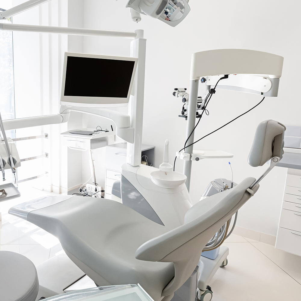
A digital impression is a detailed, three-dimensional record of your teeth and surrounding oral tissues captured with a small, wand-like intraoral scanner. Instead of using impression trays and alginate putty, the scanner records a rapid sequence of images that specialized software stitches together into an exact virtual model. That model can be viewed from any angle on a computer screen, adjusted in real time, and exported to other digital systems for design and fabrication.
For patients, the experience is noticeably different from traditional impressions: there’s no bulky material in the mouth, no gagging from tray-overflow, and no long waiting while material sets. Clinicians benefit from immediate visual feedback; if an area hasn’t been captured clearly, it can be rescanned right away. The result is a precise digital file that replaces physical molds in many restorative, orthodontic, and prosthetic workflows.
The underlying technology combines high-resolution optics and software algorithms to produce consistent results across a wide range of clinical situations. As the scanner moves through the mouth it interprets surface geometry, color, and texture, delivering a lifelike representation that supports both diagnostic review and restorative planning. This makes digital impressions a powerful tool for modern dentistry without requiring patients to have any specialized knowledge of the process.
One of the most immediate advantages of digital impressions is comfort. Traditional impressions often cause discomfort, anxiety, or a gag reflex for some patients because of the tray and impression material. By contrast, intraoral scanning is minimally intrusive: the wand is small, the process is quiet, and most patients describe it as simply a little movement in the mouth.
Beyond comfort, digital impressions streamline appointments. Captures are faster and can be completed in a single visit without waiting for materials to set or for physical models to be fabricated. For patients who have difficulty sitting for long periods, the shorter scanning time and the ability to pause and rescan targeted areas make treatment planning less taxing and more efficient.
The digital files also reduce the chance of remakes that would require additional visits. When a conventional impression contains air bubbles, distortions, or incomplete detail, the lab may reject it and request a new impression—necessitating another appointment. With digital scanning, clinicians and technicians can confirm capture quality immediately and correct any gaps while the patient is still in the chair.
Digital impressions change how the clinical team and dental laboratory interact. Once a scan is complete, the digital file can be securely transmitted to a dental technician or in-house milling unit, eliminating the delay and variability of shipping physical impressions. This direct transfer accelerates the treatment timeline and improves communication, since technicians can annotate files, request additional views, or simulate restorations before fabrication begins.
Integration with CAD/CAM systems allows dentists to design restorations virtually and, where available, manufacture them in-office the same day. For practices equipped with chairside milling or printing technology, this means a patient who needs a crown or onlay may receive a permanent restoration in a single appointment. Even when the work is sent to an external lab, the digital workflow often shortens turnaround and reduces manual steps that introduce error.
From a practice management perspective, digital impressions reduce physical storage needs and simplify record-keeping. Digital archives are searchable and easily backed up, helping maintain continuity of care and enabling efficient case retrieval for follow-up appointments or future reference.
High-quality digital impressions rival—and in many cases exceed—the precision of traditional stone models. The level of detail captured is well suited for crowns, bridges, veneers, implant prosthetics, and orthodontic appliances. For implant cases, digital files can be combined with CBCT data to plan restorations and prosthetic components with predictable fit and occlusion.
Because the digital model is manipulable, clinicians can perform virtual margin checks, occlusal adjustments, and esthetic previews before committing to fabrication. This capability supports better communication with patients, allowing them to see proposed outcomes and understand the steps involved in their treatment. For complex rehabilitations or multi-unit restorations, the ability to simulate and refine designs digitally reduces surprises during placement.
Digital impressions also integrate seamlessly with aligner and orthodontic workflows. Clear aligner manufacturers and orthodontic labs commonly accept digital scans as the basis for custom appliance design, enabling a smoother transition from diagnosis to active treatment without needing an intermediate physical model.
It’s important to note that while digital scanning is highly versatile, clinical judgment determines the best approach for each case. The practice will select the capture method that offers the most reliable outcome based on factors such as access, soft-tissue management, and restorative complexity.
During a digital impression appointment, your dental team will prepare the area by ensuring cleanliness and dryness; this helps the scanner obtain consistent data. The clinician will then guide the intraoral scanner along the teeth and soft tissues, typically in a methodical sequence to ensure full-arch or quadrant coverage. You may be asked to move your tongue or swallow at times, but the process is generally quick and well tolerated.
After the scan, the clinician reviews the 3D model on a monitor with you. Any missing detail can be rescanned immediately, which reduces the need for follow-up appointments. If a restoration is being planned, the team will discuss the next steps—whether that involves in-office fabrication, collaboration with a dental lab, or scheduling for placement once the restoration is ready.
For patients interested in same-day ceramic restorations, digital impressions are the cornerstone technology that makes single-visit treatment possible. For other treatments, the digital record speeds communication and helps ensure that the final result fits comfortably and performs as intended.
If you have concerns about sensitivity, gag reflexes, or sitting for extended periods, let the clinical team know before the scan begins. They can adjust their technique or take short breaks to maintain your comfort throughout the appointment.
In summary, digital impressions represent a comfortable, accurate, and efficient alternative to traditional impression techniques. By capturing precise digital models, the technology streamlines clinical workflows, enhances communication with dental laboratories, and supports a wide range of restorative and orthodontic treatments. If you’d like to learn how digital impressions are used at Suss Dental Group in Bergenfield, contact us for more information.


A digital impression is a high-resolution, three-dimensional record of the teeth and surrounding oral tissues captured with an intraoral scanner. The scanner records a rapid sequence of images as it moves through the mouth and specialized software stitches those images into an exact virtual model. That model can be rotated, measured, and evaluated on a computer screen to support diagnosis and restorative planning.
Digital impressions replace the need for traditional trays and putty by producing a precise digital file that can be exported to laboratory partners or CAD/CAM systems. Clinicians can review capture quality immediately and rescan any area that requires refinement while the patient is still in the chair. This immediate feedback reduces the chance of remakes and improves communication between the dental team and the laboratory.
Digital impressions eliminate bulky impression trays and the setting time required by conventional materials, which many patients find uncomfortable. The digital workflow reduces errors associated with air bubbles, distortion, or improper tray seating that can occur with physical impressions. In many cases, the level of detail captured by a modern scanner equals or exceeds that of traditional stone models for crowns, bridges, and prosthetics.
From a laboratory perspective, digital files streamline communication and reduce shipping delays because scans are transmitted electronically. Digital archives are searchable and easily backed up, simplifying record keeping and retrieval for future treatment. That said, clinical judgment determines whether a digital or conventional approach is best for any particular case based on access and soft-tissue management.
Most patients report that intraoral scanning is less invasive and better tolerated than conventional impressions because the wand is small and the process is quick and quiet. There is no impression material to trigger gag reflexes or leave an unpleasant taste, and clinicians can pause or rescan targeted areas to accommodate comfort needs. The noncontact nature of some scanners also reduces direct manipulation of soft tissues during capture.
Digital impressions do not introduce additional safety risks beyond standard dental procedures and are suitable for many patients, including those with sensitive gag reflexes or limited mouth opening. If a patient has specific concerns about sensitivity or sitting for extended periods, the clinical team can adjust technique or break the scan into shorter segments. The result is a patient-centered approach that prioritizes both comfort and clinical accuracy.
Digital impressions are widely used for single crowns, fixed bridges, veneers, inlays, onlays, and implant prosthetics because they capture fine detail necessary for precise fit. They are also integral to orthodontic workflows, providing the digital bases required by clear aligner manufacturers and orthodontic labs. In addition, digital scans support diagnostic records, occlusal analysis, and esthetic planning through virtual simulations.
For same-day dentistry, digital impressions pair with in-office milling and printing to enable single-visit restorations when the practice has chairside fabrication capabilities. Even when laboratory fabrication is needed, digital files accelerate turnaround and reduce manual steps that can introduce error. The versatility of digital scans makes them a foundational tool across restorative and orthodontic disciplines.
Yes, digital impressions are commonly combined with CBCT data for comprehensive implant planning and prosthetic design, allowing clinicians to coordinate implant position with the final restoration. The digital workflow supports virtual margin checks, emergence profile assessment, and component selection to enhance predictable fit. Implant libraries and digital analogs can be used in the design phase to ensure compatibility with restorative components.
When properly captured, digital scans produce accurate models for definitive implant prostheses and can streamline laboratory communication for screw-retained or cement-retained restorations. Complex multi-unit rehabilitations benefit from the ability to simulate occlusion and esthetic outcomes before fabrication. Clinical judgment remains essential to determine when adjunctive techniques or traditional impressions may be warranted to manage soft tissue or access issues.
Digital impressions often shorten treatment timelines by allowing immediate digital transfer of files to laboratories or in-office CAD/CAM systems, eliminating shipping delays associated with physical models. Technicians can review scans, request additional views, or begin designing restorations the same day the scan is captured. For practices with chairside milling or printing equipment, a final restoration can sometimes be delivered in a single visit.
Beyond speed, the digital workflow reduces manual steps and potential sources of error during model fabrication, which improves consistency and predictability. Digital archiving simplifies case retrieval for follow-up care and long-term record keeping. The result is a more efficient practice workflow that benefits both clinicians and patients without compromising clinical standards.
Before scanning begins, the clinician will prepare the area by ensuring cleanliness and relative dryness to help the scanner capture consistent data. The scan itself involves a methodical pass of the intraoral wand over the teeth and soft tissues, during which the patient may be asked to move the tongue or pause briefly for specific views. The process is typically quick and well tolerated, and most scans can be completed in a single visit for the area being treated.
After capture, the clinician reviews the 3D model on a monitor and can immediately rescan any missing or unclear areas, reducing the need for repeat visits. The dental team will then discuss the next steps for restoration design, fabrication, or scheduling. Patients interested in same-day restorations should ask whether chairside fabrication is available at their practice.
High-quality digital impressions provide excellent accuracy for many single-unit and multi-unit restorations when captured with proper technique and equipment. Scanners interpret surface geometry, color, and texture to produce lifelike representations that support margin verification and occlusal analysis. For lengthy spans or full-arch cases, clinicians follow specific scanning protocols and may use adjunctive techniques to maintain precision across the arch.
For complex rehabilitations, the digital model can be combined with other imaging modalities and virtual planning tools to predict fit and esthetic outcomes. While digital systems continue to improve, clinical judgment guides whether a digital approach alone is appropriate or whether supplemental data or traditional techniques are needed. Collaboration with the laboratory and careful case planning enhance predictability in complex situations.
Digital impressions are highly versatile, but certain clinical situations may challenge scanner access or soft-tissue visualization, such as deeply subgingival margins or limited mouth opening. In cases with heavy bleeding, excessive saliva, or poor access, capturing a consistent digital scan can be more difficult and may require soft-tissue management or alternative techniques. Some clinicians may still choose conventional impressions for specific laboratory workflows or when working with materials that require physical models.
Decisions about capture method are based on case complexity, restorative goals, and clinician experience rather than a one-size-fits-all rule. When a digital scan is used, proper isolation and technique are important to achieve the best result. The practice will select the approach that provides the most reliable outcome for each patient.
Suss Dental Group employs intraoral scanning as a core part of restorative and orthodontic workflows to improve accuracy, comfort, and communication with laboratories. Scans are reviewed with patients on-screen to explain treatment options and to show how proposed restorations or aligner outcomes will look. When chairside fabrication is available, digital impressions enable same-day ceramic restorations; when laboratory fabrication is needed, digital files accelerate coordination and reduce turnaround.
The practice combines scanner technology with clinical judgment to select the most appropriate capture method for each case, ensuring reliable results across routine and complex treatments. Patients are encouraged to discuss comfort concerns or specific clinical needs with the dental team so scanning can be adapted as necessary. For those interested in learning more, contacting Suss Dental Group in Bergenfield will provide information about how digital impressions are used in individual treatment plans.
Ready to schedule your next dental appointment or have questions about our services?
Contacting Suss Dental Group is easy! Our friendly staff is available to assist you with scheduling appointments, answering inquiries about treatment options, and addressing any concerns you may have. Whether you prefer to give us a call, send us an email, or fill out our convenient online contact form, we're here to help. Don't wait to take the first step towards achieving the smile of your dreams – reach out to us today and discover the difference personalized dental care can make.